Left Atrial Appendage Closure Device Program
What is a Left Atrial Appendage and Why Does it Need Closing?
 Left Atrial Appendage Closure Device
Most people are not aware that they have a left atrial appendage (LAA). The LAA is a small sac or pouch, connected to the top left side of your heart (the left atrium). Each person's LAA is different in size and shape. Most do not experience issues with their LAA; however, when an individual has atrial fibrillation (AFib) and their heart is not pumping blood as it should, blood can collect in the LAA and form clots. If a clot escapes, it can cut off the blood supply to the brain – causing a stroke. 90% of stroke-causing blood clots that come from the heart are formed in the LAA.
The left atrial appendage closure device reduces stroke risk in people with the type of atrial fibrillation (AFib) not caused by a heart valve problem. It works differently from blood thinners.
How the Left Atrial Appendage Closure Device Works
- The left atrial appendage closure device fits into a part of your heart called the left atrial appendage (LAA).
- The left atrial appendage closure device permanently closes off this part of your heart to keep those blood clots from escaping.
The Left Atrial Appendage Closure Device Procedure
The left atrial appendage closure device is about the size of a quarter. Implantation does not require open-heart surgery. Here is what happens during the procedure.
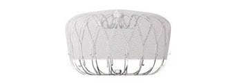
 Boston Scientific Watchman™
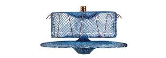
 Abbott Amplatzer™ Amulet™
- To implant the device, the doctor makes a small cut in your upper leg and inserts a narrow tube, called a catheter.
- The doctor then guides the implant through the catheter, into your left atrial appendage (LAA).
- The procedure is performed under general anesthesia and typically takes about an hour. People who have this device implanted may be discharged to home the same day or stay overnight, depending on physician direction.
- Heart tissue gradually grows over the closure device to form a barrier against blood clots.
To determine your eligibility for device implant, you may need the following:
Transesophageal Echocardiogram (TEE)
An echocardiogram (echo) test is when ultrasound or sound waves are used to make an image of your heart. During a transesophageal echo (TEE) a physician inserts a narrow tube with a small probe into your mouth and down your esophagus (throat). This allows the doctor to record images of your heart from the inside, providing images closer to your heart. The physician will then use the information collected during the TEE to determine which device is the most appropriate.
Coronary Computed Tomography Angiogram (CCTA) Only if Necessary
You will lie on a table that moves into a scanner. The scanner will take detailed pictures of your blood vessels. This will tell the doctor if the blood vessels in your legs are big enough for the catheter that carries your new valve to your heart. This test often includes a dose of contrast (X-ray) dye into a vein in your arm. We may change this test for you if your kidneys don’t work well.