Vascular Surgery Procedures & Interventions
Abdominal Aortic Aneurysm Stent Grafting and Surgical Repair
Endovascular Abdominal Aortic Aneurysm Repair (EVAR) is a procedure that repairs an abdominal aortic aneurysm (AAA), by placing a stent graft under X-ray through two small access sites in both groins. The stent graft is a long, mesh tube that is placed in the abdominal aorta, redirecting blood flow away from the aneurysm and to the graft, decreasing pressure on the aneurysm. An AAA also can be repaired surgically, with open abdominal aortic aneurysm repair. This is done in the operating room through a large incision over the abdomen.
%20abdominal%20aortic%20aneurysm%20(AAA).png)
Angiograms with Angioplasty, Stents and Thrombectomy
An angiogram/aortogram can be described as an X-ray of the blood vessels (more specifically the arteries) using contrast to evaluate blood flow. This procedure is done when we suspect an issue with blood flow through the body or to an extremity. A catheter is usually placed through a small access site, most commonly in the groin or arm. To improve the blood flow, balloon angioplasty, stent placement, thrombectomy or thrombolysis can be used.
- When angioplasty is performed a balloon is generally used in the artery and inflated to open up any narrowing (or stenosis) in the artery.
- A stent also can be placed in areas where the artery is narrowed or where a balloon is not sufficient. The stent provides a support structure to keep the artery open.
- Thrombectomy or thrombolysis is used when a blood clot (thrombus) is found in an artery. The doctor injects clot-busting medication directly in the location of the clot and sucks out as much of the clot as possible to restore blood flow in the area.
Embolectomy
An embolectomy is surgery to remove an embolus located in an artery or vein. An embolus is part of a blood clot that has broken free from the original clot. The removal of embolus is important as it can move and become stuck in another area, blocking blood flow or putting you at risk for a stroke. There are three options for performing an embolectomy: catheter, balloon and open.
- A catheter embolectomy is when a catheter (or thin tube) is guided into the vein or artery that has the embolus. Suction is used to remove the embolus through the catheter.
- A balloon embolectomy is when a catheter with a (deflated) balloon is guided into the vein, past the clot. The balloon is inflated and pulled back, taking the embolus back through the vein with the catheter. The balloon helps move the embolus and keep it from continuing into the vein.
- An open embolectomy usually is only done for a large embolus, most often to treat a pulmonary embolism (PE). Your surgeon will make an incision in the skin over the embolus, open the vein or artery and remove the embolus.
Endarterectomy
An endarterectomy is a procedure to remove plaque buildup inside an artery. Plaque buildup often is found in the carotid and femoral arteries. An endarterectomy helps improve blood flow and relieve symptoms caused by a narrowed or blocked artery.
- Carotid arteries are the main blood vessels that carry blood and oxygen to the brain. Removing plaque in this area is known as a carotid endarterectomy.
- Femoral arteries are the main blood vessels in your thighs that carry blood and oxygen to your legs. Removing plaque in this area is known as a femoral artery endarterectomy.
Arterial Bypasses
Arterial bypasses are performed to create a new route for blood to flow around a blocked portion of an artery.
-
- Femoral-popliteal bypass is a surgery to place a graft that goes around narrowed arteries in your upper leg, improving blood flow to your leg and foot.
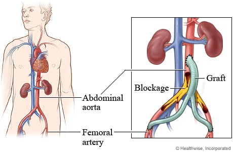
- Aorto-femoral bypass is surgery to place a graft that goes around your blocked or damaged aorta. The aorta is a large blood vessel that carries blood and oxygen from your heart to your body. Your aorta splits into two smaller blood vessels called femoral arteries in your abdomen. These arteries carry blood and oxygen to your pelvis and legs.

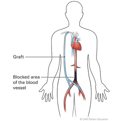
- Axillofemoral bypass is a surgery to place a graft around your blocked or damaged aorta, going around (bypassing) the blocked arteries of the abdomen and rerouting the blood by connecting your axillary artery (located in your shoulder) to the femoral artery in your groin/legs.

Arteriovenous (AV) Fistula Placement
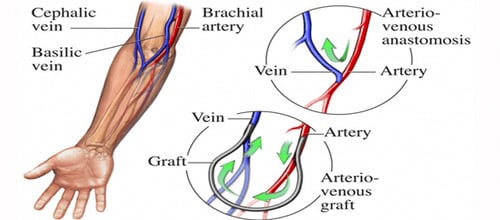
An AV fistula creation is surgery to connect an artery to a vein. This surgery is done so you can receive hemodialysis. The AV fistula is usually placed in your forearm or upper arm.

An AV graft creation is surgery to connect an artery to a vein using a graft (tube). You may need an AV graft if your artery and vein cannot be directly joined together for hemodialysis. The AV graft is usually placed in your forearm or upper arm.

Fistulagram
A fistulagram is a procedure that looks at the blood flow and checks for blood clots, blockages or narrowing of an artery or vein in your fistula. Blood clots, blockages, or narrowing may interfere with your dialysis treatments.
Carotid Artery Stenting and Surgery
Carotid arteries in your neck supply oxygen and blood to your brain. Stenosis is when plaque builds up inside the arteries. This plaque causes arteries to narrow which restricts blood flow. If plaque becomes severe it could cause a stroke.
-
- Carotid endarterectomy (CEA) is a surgery performed to remove plaque (fatty deposits) from inside your carotid artery. The carotid artery is a blood vessel found in both sides of your neck. Plaque may build up inside your carotid artery and decrease blood flow to your brain. A piece of plaque could also break free and cause a stroke
.png)
- Transcarotid Artery Revascularization (TCAR) procedure is typically reserved for high-risk surgical patients.
- Carotid artery stent placement is a procedure to widen a narrowed carotid artery. The carotid arteries are two large blood vessels on each side of your neck. They carry blood and oxygen from your heart to your brain. A stent is a wire mesh tube that helps hold your carotid artery open.
Percutaneous Thrombectomy for Pulmonary Embolism (PE)
One of the most serious complications of Deep Vein Thrombosis (DVT) is a pulmonary embolism (PE). A pulmonary embolism occurs when the clot breaks loose from the vein wall and travels to the lungs, blocking the artery that supplies blood to the lungs. Percutaneous thrombectomy is a method of retrieving the embolism by using special catheters that can break it loose. Suction is used to pull out the PE through the catheter, removing it from it the artery.
Compartment Syndrome
Compartment syndrome is a painful condition that occurs when pressure under the tissue of a muscle (fascia) becomes so great, it begins to cut off blood flow and nerve signals to muscles and surrounding tissues. Fasciotomy is a surgery where an incision is made in the fascia to relieve the pressure. A fasciotomy can be done on most areas of the body, but is most common on the arm or leg.
Deep Vein Thrombosis (DVT) Treatment
Deep Vein Thrombosis (DVT) occurs when a blood clot forms in a deep vein, usually in the leg. DVT also can occur in other places, such as your arm or chest. Most of these clots occur when blood flow in the vein is slowed, usually because of inactivity. Normally, as you walk around your leg muscles squeeze your veins and keep blood flowing back to the heart. But if you are inactive for many hours – such as during a long airplane flight or while recovering from an operation or stroke – blood flow in the veins may slow so much that a blood clot forms.
- Mechanical thrombectomy is an emergency procedure used to remove a blood clot from a blood vessel (vein or artery). A clot that breaks free and travels to the lungs can cause a pulmonary embolism (PE). Even if the clot does not break free, it can prevent blood flow to body areas.
- Anticoagulants are blood thinning medications that reduce the risk of blood clots.